NCERT Class 11 Biology Chapter 8 Notes Cell The Unit Of Life- Download PDF Notes
Have you ever wondered what makes living things different from non-living ones? It’s the presence of cells, which are the tiny building blocks that give life to every organism. The NCERT Class 11 Biology Chapter 8 Notes Cell The Unit of Life cover every topic in a well-understandable language with well-labelled diagrams. Important definitions and terms are highlighted for better understanding by the students. This chapter includes the cell theory, types of cells, different cell organelles, and their functions. Students can use the NCERT Notes to gain clarity before board exams as well as entrance exams like NEET.
This Story also Contains
- NCERT Class 11 Biology Chapter 8 Notes: Download PDF
- Class 11 Biology Chapter 8 Cell: The Unit of Life Notes
- Cell: The Unit of Life Previous Year Questions and Answers
- How to Use the Cell: The Unit of Life Class 11 Notes Effectively?
- Advantages of Class 11 Biology Chapter 8 Cell The Unit of Life Notes
- Chapter-Wise NCERT Class 11 Notes Biology

The cell is the basic structural and functional unit of life because it carries out all life processes in the body. Cell: The Unit of Life Class 11 Notes covers topics like the types of cells, differences between prokaryotic and eukaryotic cells, cell theory, and cell organelles. Students can also download the PDF of Cell The Unit of Life Class 11 Notes for quick revision. Using these NCERT Notes for Class 11 regularly helps students understand important topics, which makes them feel more confident during the exam.
NCERT Class 11 Biology Chapter 8 Notes: Download PDF
Students can easily download the complete notes for the chapter Cell: The Unit of Life in PDF format. These notes are great for quick revision and last-minute study. Students can use Cell The Unit of Life Class 11 Notes for offline revision. The PDF helps students understand and remember topics faster. The NCERT Class 11 Biology Notes are a valuable resource for understanding complex topics in a simple way.
Also, students can refer to:
Class 11 Biology Chapter 8 Cell: The Unit of Life Notes
Students can understand the structure and function of cells by using these notes. Important cell organelles like the nucleus, mitochondria, Golgi apparatus, and ribosomes are described in clear points. Important terms of this chapter help students understand the concepts easily without any further help. All topics are covered in the Cell: The Unit of Life Class 11 Notes in such a way that they follow the latest NCERT curriculum.
Cell Theory
Matthias Schleiden, a German botanist, researched plants in 1838 and discovered that all plants consist of various forms of cells, which collectively constitute plant tissues.
At about the same period, British zoologist Theodore Schwann (1839) examined animal cells and found that they have a thin outer covering, termed the plasma membrane. He also described that plant cells possess a cell wall that is not found in animal cells.
Schleiden and Schwann collectively developed the cell theory.
But their theory didn't account for the way in which new cells are created. In 1855, Rudolf Virchow expanded on the theory by declaring that new cells come from existing cells.
Modern Cell Theory Holds That:
- All living things consist of cells and their byproducts.
- New cells are produced only from existing cells via cell division.
Prokaryotic Cells
Prokaryotic cells are tiny compared to eukaryotic cells and reproduce rapidly. They are also available in various shapes, which are as follows:
- Bacillus (rod-shaped)
- Coccus (spherical)
- Vibrio (comma-shaped)
- Spirillum (spiral-shaped)
Even though prokaryotic cells differ from each other in terms of shape and functionality, they follow a common basic structure. All prokaryotes except Mycoplasma have a cell wall outside the cell membrane.
The cytoplasm is a jelly-like liquid within the cell. Prokaryotes do not have a distinct nucleus since their genetic material is not covered with a nuclear membrane. Rather, their DNA occurs freely in the cytoplasm.
Plasmids and Genetic Material
- Plasmids occur in many bacteria as small circular DNA fragments beyond their primary DNA.
- Plasmids provide bacteria with unique characteristics, including antibiotic resistance.
- Researchers employ plasmids in genetic engineering to introduce foreign DNA into bacteria.
- In contrast to eukaryotic cells, prokaryotic cells do not have membrane-bound organelles other than ribosomes.
Cell Structure and Envelope
Prokaryotic cells possess a complex protective layer known as the cell envelope, which is composed of:
- Glycocalyx (outer layer) – May be a slime layer (loose covering) or a capsule (tough outer coating).
- Cell Wall (middle layer) – Gives strength and support.
- Plasma Membrane (inner layer) – A phospholipid bilayer that protects the cell and facilitates communication.
- Bacteria are differentiated as Gram-positive or Gram-negative by their cell wall structure, by the method known as Gram staining, invented by Hans Christian Gram.
Movement and Surface Structures
Flagella and cilia facilitate movement, propelled by a basal body.
Pili and fimbriae are short, hair-like structures facilitating surface attachment for bacteria.
Internal Structures
- Mesosomes – Folding of the cell membrane that helps in cell wall construction and replication of DNA.
- Chromatophores – Present in photosynthetic bacteria such as cyanobacteria, they contain pigments for photosynthesis.
- Ribosomes – Small organelles in the cytoplasm that assist in protein synthesis. They are categorised according to their rate of sedimentation in Svedberg (S) units.
Eukaryotic Cells
Eukaryotic cells are larger and more complex than prokaryotic cells. They have a well-defined nucleus enclosed by a nuclear membrane and contain membrane-bound organelles that perform specialised functions. Eukaryotic cells can be unicellular (like amoebas) or multicellular (like plants and animals).
Cell Structure
Eukaryotic cells have three main parts:
Plasma Membrane – A phospholipid bilayer that protects the cell and controls what enters and exits.
Nucleus – The control centre of the cell, containing DNA and responsible for growth and reproduction.
Cytoplasm – A fluid-filled space containing organelles that carry out life functions.
Cell Organelles and Their Functions
Nucleus – Contains chromosomes (DNA) and directs cell activities.
Endoplasmic Reticulum (ER) – Helps in protein and lipid synthesis.
Rough ER has ribosomes and makes proteins.
Smooth ER makes lipids and detoxifies chemicals.
Golgi Apparatus – Packages and transports proteins and lipids.
Mitochondria – The powerhouse of the cell, producing energy (ATP) through cellular respiration.
Lysosomes – Contain digestive enzymes to break down waste.
Plastids (only in plants) – Help in photosynthesis (chloroplasts) and store food (leucoplasts).
Vacuoles – Store water, nutrients, and waste; larger in plant cells.
Cell Wall and Cytoskeleton
Cell Wall (only in plant cells) – Provides structure and support.
Cytoskeleton – A network of protein filaments that maintains cell shape and helps in movement.
Differences Between Plant and Animal Cells
Plant Cells have a cell wall, a large vacuole, and chloroplasts for photosynthesis.
Animal Cells have centrioles and lysosomes, which help in cell division and digestion.
Eukaryotic cells allow complex life forms to exist by efficiently carrying out essential biological functions through their specialised organelles.
Plant Cell VS. Animal Cell
- Plant cells have a cell wall and chloroplasts, which help in photosynthesis, while animal cells do not.
- Plant cells have a large central vacuole, whereas animal cells have small or no vacuoles.
- Both have a nucleus, cytoplasm, and cell membrane.
- Animal cells are generally round, and plant cells are rectangular due to the rigid cell wall.
- Plant cells also store energy as starch, while animal cells store it as glycogen.
Cell Organelles
Cell organelles are specialised structures contained within a cell that have specific functions to maintain the cell alive and in its optimum working condition. They are membrane-bound in eukaryotic cells, while prokaryotic cells have most of them missing, except for ribosomes. Here is a detailed description of each organelle and what it does.
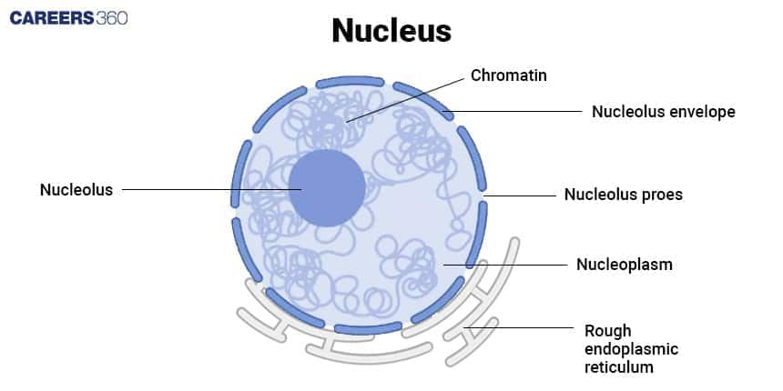
Nucleus – The Control Centre
The nucleus is the largest organelle in eukaryotic cells.
It contains genetic material (DNA), which controls cell activities.
The nuclear membrane encloses the nucleus and has pores for the exchange of materials.
Inside the nucleus, the nucleolus is responsible for making ribosomes.

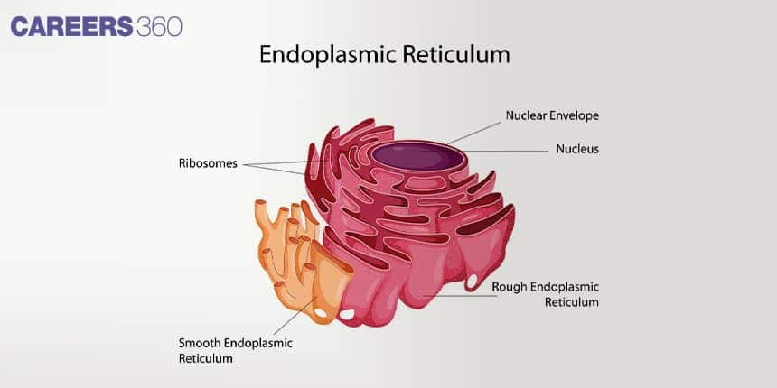
Endoplasmic Reticulum (ER) – The Transport Network
A network of membranes that transports materials within the cell.
Types:
Rough ER (RER): Studded with ribosomes, it helps in protein synthesis.
Smooth ER (SER): Lacks ribosomes, produces lipids, and detoxifies harmful substances.

Ribosomes – The Protein Factories
Tiny structures are responsible for protein synthesis.
Found freely floating in the cytoplasm or attached to the rough ER.
Present in both prokaryotic and eukaryotic cells.
Golgi Apparatus – The Packaging and Dispatch Centre
A stack of flattened sacs that modifies, sorts, and packages proteins and lipids.
The Golgi Apparatus sends these materials to their destinations within or outside the cell.
Produces lysosomes in animal cells.
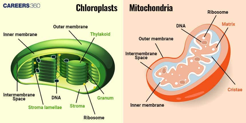
Mitochondria – The Powerhouse of the Cell
Mitochondria produce energy (ATP) through cellular respiration.
They have a double membrane, with the inner membrane forming folds called cristae to increase surface area for energy production.
They contain their own DNA and ribosomes, allowing them to reproduce independently.

Lysosomes – The Digestive System of the Cell
Small vesicles filled with enzymes that break down waste materials, old cell parts, and foreign substances.
Help in cell defence by destroying harmful invaders.
Known as suicidal bags, as they can cause cell self-destruction if necessary.
Vacuoles – Storage Units
Fluid-filled sacs are used for storing water, nutrients, and waste.
Large and central in plant cells to provide support.
Smaller in animal cells and helps in waste disposal.
Plastids – The Colour and Food Factory (Only in Plant Cells)
Chloroplasts – Contain chlorophyll and perform photosynthesis.
Chromoplasts – Provide pigments to flowers and fruits.
Leucoplasts – Store starch, proteins, and fats.
Cytoskeleton – The Support System
A network of protein filaments that gives the cell its shape and structure.
Helps in cell movement and internal transport.
Peroxisomes – The Detox Centres
Contains enzymes that break down fatty acids and harmful substances like hydrogen peroxide.
Help in the detoxification of the cell.
Cilia and Flagella – The Movement Organelles
Cilia – Small, hair-like structures that help in movement and sensing the environment.
Flagella – Long, whip-like structures that help in cell movement, seen in sperm cells and bacteria.
Centrosome and Centrioles – The Cell Division Helpers (Only in Animal Cells)
The centrosome contains centrioles, which help in cell division by forming the spindle fibers during mitosis.
Also, Read
Cell: The Unit of Life Previous Year Questions and Answers
Here are important questions from past papers based on this chapter to help students understand the level of difficulty. To solve these questions without any difficulty, students can use the NCERT Class 11 Biology Chapter 8 Notes.
Question 1. Who proposed the fluid-mosaic model of the plasma membrane?
Option 1. Camillo Golgi
Option 2. Schleiden and Schwann
Option 3. Singer and Nicolson
Option 4. Robert Brown
Answer :
An important turning point in the study of cellular organelles was reached in 1898 when Camillo Golgi discovered the Golgi Complex. All living things are made up of cells, which are the basic building blocks of life, according to the Cell Theory, which was put forth by Matthias Schleiden and Theodor Schwann in 1839. In the same vein, Robert Brown identified the nucleus as an essential part of plant cells in his groundbreaking study from 1831, which gave the first thorough description of the organelle.
Hence, the correct answer is option (3), Singer and Nicolson
Question 2. A common characteristic feature of plant sieve tube cells and most mammalian erythrocytes is
Option 1. Absence of mitochondria
Option 2. Presence of a cell wall
Option 3. Presence of haemoglobin
Option 4. Absence of a nucleus
Answer:
A common characteristic feature of plant sieve tube cells and most mammalian erythrocytes is the absence of a nucleus at the time of maturity. In sieve tube cells, this special adaptation maximises space for the efficient transport of sugars and nutrients within the phloem. Similarly, the erythrocytes of mammals lose their nucleus during maturation to provide more space for hemoglobin, optimising their work of carrying and delivering oxygen throughout the body.
Hence, the correct answer is option (4) Absence of nucleus
Question 3. Which of the following is not true of a eukaryotic cell?
Option 1. The cell wall is made up of peptidoglycans
Option 2. It has an 80S-type ribosome present in the cytoplasm
Option 3. Mitochondria contain circular DNA
Option 4. Membrane-bound organelles are present
Answer:
The main building block of the cell walls of eukaryotic plant cells is cellulose, a structural polysaccharide. It supports the cell's structure and shields it from mechanical stress from the outside by giving it strength and stiffness.
Hence, the correct answer is option (1), the Cell wall is made up of peptidoglycans
Question 4. Which organelle is responsible for the detoxification of drugs and poisons in eukaryotic cells?
Option 1. Rough Endoplasmic Reticulum
Option 2. Smooth Endoplasmic Reticulum
Option 3. Golgi apparatus
Option 4. Lysosomes
Answer:
The Smooth Endoplasmic Reticulum (SER) is involved in lipid synthesis and plays a major role in the detoxification of drugs and harmful chemicals, especially in liver cells. It contains enzymes that modify and neutralise toxic substances, making them easier for the body to eliminate.
Hence, the correct answer is option (2) Smooth Endoplasmic Reticulum
Question 5. Which of the following structures is not found in prokaryotic cells?
Option 1. Ribosomes
Option 2. Nucleoid
Option 3. Plasma membrane
Option 4. Mitochondria
Answer:
Prokaryotic cells have a nucleoid, ribosomes, and a plasma membrane, but they lack membrane-bound organelles such as mitochondria, chloroplasts, Golgi bodies, and ER. Since mitochondria are membrane-bound, they are absent in all prokaryotes, including bacteria and archaea.
Hence, the correct answer is option (4) Mitochondria
Also Read:
How to Use the Cell: The Unit of Life Class 11 Notes Effectively?
Clear and well-arranged notes can make the chapter easier to understand and revise during exams. Given below are a few ways by which students can make the most use of the notes.
Focus on understanding the differences between prokaryotic and eukaryotic cells, as the difference is frequently asked in exams.
Go through Class 11 Biology Chapter 8 Cell: The Unit of Life Notes to revise definitions like cell theory, plasma membrane models, and cell organelles.
Make flowcharts for different processes to understand them easily without wasting much time.
Highlight the unique features of each organelle (like mitochondria as the powerhouse, ribosomes as protein factories) while revising.
Use the Class 11 Biology Chapter 8 Cell The Unit of Life Notes PDFs to connect theory with diagrams, since visual learning is important for both board and NEET exams.
Advantages of Class 11 Biology Chapter 8 Cell The Unit of Life Notes
The notes help students build a strong foundation in understanding the structure and function of cells and cell organelles. Going through the notes offers many advantages to students, some of which are listed below:
- Class 11 Biology Chapter 8 Cell The Unit of Life Notes PDF provides a summary of cell theory, types of cells, and cell organelles in an easy-to-understand manner.
- The notes include clear and well-labeled diagrams, by which students can improve their visual learning.
- All the functions and the structure of important cell organelles like mitochondria, nucleus, and ribosomes are explained, which are frequently asked in exams.
- Students can go through these notes when they have less time for doing a quick and easy revision.
Chapter-Wise NCERT Class 11 Notes Biology
Given below is a complete set of notes covering all chapters in Class 11 Biology. They include important definitions, diagrams, and important points for exam preparation.
Frequently Asked Questions (FAQs)
The nucleus stores genetic material (DNA), regulates gene expression, and controls cellular activities. It contains the nucleolus, which is responsible for ribosome synthesis.
Mitochondria generate ATP through cellular respiration, providing energy for cellular functions. They contain their own DNA, enabling self-replication and energy production.
The nucleus stores genetic material (DNA), regulates gene expression, and controls cellular activities. It contains the nucleolus, which is responsible for ribosome synthesis.
Cells are categorized into prokaryotic (bacteria, archaea) and eukaryotic (plants, animals, fungi, protists) as mentioned in the NCERT Class 11 Biology Chapter 8 Notes Cell The Unit of Life. Prokaryotic cells lack a true nucleus and membrane-bound organelles, whereas eukaryotic cells possess a well-defined nucleus and organelles.
Eukaryotic cells consist of a plasma membrane, nucleus, cytoplasm, and organelles like mitochondria, ribosomes, endoplasmic reticulum, Golgi apparatus, lysosomes, and vacuoles, each contributing to cell function and maintenance. These are well-explained in the NCERT Class 11 Biology Chapter 8 Notes Cell The Unit of Life.
A cell is the fundamental structural and functional unit of life. It is the smallest unit capable of independent existence and performing essential life processes. Cells can be unicellular or multicellular, varying in structure and function.
Prokaryotic cells are smaller, lack a nucleus, and contain circular DNA. They do not have membrane-bound organelles. Eukaryotic cells are larger, have a nucleus with linear DNA, and contain organelles like mitochondria, ER, and Golgi apparatus.
The plasma membrane regulates the entry and exit of substances, maintaining homeostasis. It is selectively permeable, allowing nutrients, gases, and waste to pass through while protecting the cell from external environments.
Popular Questions
Courses After 12th
Applications for Admissions are open.
As per latest syllabus. Physics formulas, equations, & laws of class 11 & 12th chapters
JEE Main Important Chemistry formulas
Get nowAs per latest syllabus. Chemistry formulas, equations, & laws of class 11 & 12th chapters
JEE Main high scoring chapters and topics
Get nowAs per latest 2024 syllabus. Study 40% syllabus and score upto 100% marks in JEE
JEE Main Important Mathematics Formulas
Get nowAs per latest syllabus. Maths formulas, equations, & theorems of class 11 & 12th chapters
