

NCERT Class 11 Biology Chapter 20 Notes Locomotion And Movement- Download PDF Notes
Did you know that humans have more than 600 muscles that help in movement and support? The NCERT Class 11 Biology Chapter 17 Notes Locomotion and Movement explain all the topics in a step-by-step manner. The notes are written in detail and include diagrams that make learning better. Types of movement, muscle structure, and the mechanism of muscle contraction are included. These NCERT notes help students understand the concepts quickly and clearly without any confusion.
This Story also Contains
- NCERT Class 11 Biology Chapter 17 Locomotion and Movement Notes: Download PDF
- Class 11 Biology Chapter 17 Locomotion and Movement Notes
- Chapter 17 Locomotion and Movement: Previous Year Questions and Answers
- How to Use Locomotion and Movement Class 11 Notes Effectively?
- Advantages of Class 11 Biology Chapter 17 Locomotion and Movement Notes
- Chapter-Wise NCERT Class 11 Notes Biology
Locomotion and Movement Class 11 Notes explain the role of bones and joints in simple language. Different disorders of the muscular and skeletal systems are also highlighted. The NCERT Class 11 Biology Chapter 17 Notes PDF are great for doing regular practice. Knowing these concepts is important in understanding how organisms interact with the environment and perform daily activities. The NCERT Notes for Class 11 save a lot of time while doing revision during exams.
NCERT Class 11 Biology Chapter 17 Locomotion and Movement Notes: Download PDF
To download the PDF for Locomotion and Movement, students can click on the link provided. These notes explain key topics like types of muscles, muscle contraction, and the skeletal system. The NCERT Class 11 Biology Chapter 17 Notes PDF can be accessed offline for quick learning. Diagrams and short summaries make it easy to understand and revise. The NCERT Notes for Class 11 Biology are a valuable resource for understanding the basic and difficult concepts.
Also Read,
Class 11 Biology Chapter 17 Locomotion and Movement Notes
This chapter includes topics like types of movement, the structure of muscles, and the skeletal system. The Locomotion and Movement Class 11 Notes explain the mechanism of muscle contraction in a simple way. These are designed by subject experts and follow the latest NCERT curriculum, which is helpful for quick revision and better understanding during exams.
Introduction
Movement is important for survival as it helps in searching for food, escaping predators, and reproduction in animals. Locomotion is carried out by a well-organised, muscular, and skeletal system controlled by the nervous system. Different organisms have evolved various forms of movement.
Types of Movement
The types of movement are described below-
Amoeboid:
Amoeboid movement is seen in unicellular organisms like Amoeba and in certain cells of multicellular organisms, such as leucocytes. It involves the formation of temporary projections called pseudopodia, which help in engulfing food and movement.
Ciliary:
Ciliary movement is by small hair-like structures called cilia that beat in a coordinated manner. This type of movement is seen in the respiratory tract and the reproductive tract. Paramecium, a unicellular organism, uses cilia for locomotion in water.
Muscular:
Muscular movement is the most advanced form of movement seen in higher animals, including humans. It is based on the contraction and relaxation of muscle fibres and is responsible for various functions such as walking, running, breathing, and pumping blood.
Muscle
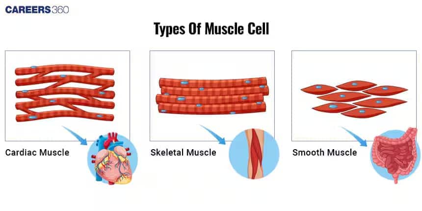
Muscles are made up of muscle fibres, which are composed of protein filaments called actin and myosin. These proteins play an important role in muscle contraction. Muscles are classified based on their structural function into three types:
Skeletal muscles: These are voluntary muscles attached to bones and help in movement.
Visceral Muscles: These are involuntary muscles found in internal organs like the stomach and intestine.
Cardiac Muscles: These are involuntary muscles found only in the heart and help in continuous contraction for pumping blood.
Structure of Skeletal Muscle
Skeletal muscle is a striated, voluntary muscle responsible for body movement. It is made up of muscle fibres, which are long, cylindrical, and multinucleate cells. These fibres are grouped into bundles surrounded by connective tissues like epimysium, perimysium, and endomysium. Each muscle fibre contains myofibrils that are further composed of repeating units called sarcomeres- the functional unit of contraction. Sarcomere contains thick (myosin) and thin (actin) filaments arranged in an overlapping pattern that gives skeletal muscles a striated appearance. Skeletal muscles are richly supplied with nerves and blood vessels, ensuring proper contraction and oxygen supply. It is attached to bones by tendons, enabling movement through contraction and relaxation.
Structure of Sarcomere
The sarcomere is the functional unit of a striated muscle fibre responsible for muscle contraction. It is composed of a thick filament arranged in a highly organised repeating pattern between two lines and consists of different bands and zones that contribute to muscle contraction. Z-lines (or Z-discs) mark the boundaries of a sarcomere and anchor the thin (actin) filaments. Thin filaments are made up of actin, troponin, and tropomyosin proteins. Actin filaments are attached to the Z line and extend to the centre of the sarcomere. Thick filaments are made up of myosin molecules with globular heads that form cross-bridges with actin during contraction. I-band(Isotropic band) is the region containing only thin filaments and shortens during muscle contraction. A-band(Anisotropic band) is the region containing the entire length of the thick filament and remains constant during contraction. The central region of the A-band contains only thick filaments called the H-zone and disappears during muscle contraction as actin slides over myosin. The M-line of the sarcomere holds the thick filament together and provides structural support to the myosin filaments.
Structure Of Contractile Protein:-
The two main contractile proteins in muscles are actin(thin filaments) and myosin(thick filaments). Actin filaments are made up of globular actin(G-actin) subunits that polymerise to form filamentous actin(F-actin). These filaments are associated with regulatory proteins like troponin and tropomyosin, which control muscle contraction and consist of myosin molecules, each with a tail and head. The myosin head has ATPase activity, which provides energy for contraction. During contraction, the myosin heads bind to actin filaments, forming cross-bridges that pull actin towards the sarcomere's centre, leading to the shortening of the muscle fibre.
Mechanism of Muscle Contraction (Sliding Filament Theory):-
A motor neuron releases the neurotransmitter acetylcholine at the neuromuscular junction.
Acetylcholine binds to the receptors on the muscle fibre, triggering an action potential across the muscle membrane.
The action potential travels through T-tubules, reaching the sarcoplasmic reticulum, which releases calcium ions into the sarcoplasm.
Calcium ions play an important role in initiating muscle contraction by binding to troponin, a protein on actin filaments.
Troponin undergoes a conformational change, causing tropomyosin to move away, exposing the myosin binding sites on actin.
The myosin heads are attached to the exposed binding sites and actin filaments, forming cross-bridges.
Myosin is in an energised state, carrying ATP that is hydrolysed into ADP and inorganic phosphate.
Once the cross-bridge is formed, myosin heads pivot, pulling the actin filament toward the centre of the sarcomere.
This action is called a power stroke, which results in muscle contraction.
A new ATP molecule binds to myosin, causing it to detach from actin, which prevents continuous binding and allows relaxation.
The ATP bound to myosin is hydrolysed, re-energising the myosin to return to its original position.
Once the nerve impulse stops, calcium ions are pumped back into the sarcoplasmic reticulum.
Troponin and Tropomyosin cover the myosin binding sites again, preventing further cross-bridge formation.
Skeletal System
Consists of a framework of bones and a few cartilages.
The human skeletal system consists of 206 bones: Axial skeleton- 80 bones, and Appendicular skeleton– 126 bones
Axial Skeleton:
The axial skeleton forms a central framework of the body, consisting of 80 bones. It includes the skull, vertebral column, ribs, and sternum. The skull has 22 bones, protects the brain, and houses sensory organs. The vertebral column has 33 vertebrae, providing structural support and flexibility and allowing movement while protecting the spinal cord. The rib cage has 12 pairs of ribs and a sternum that protect vital organs like the heart and lungs. The skeleton maintains posture, protects vital organs, and serves as an attachment for muscles involved in the movement.
Ribs- 12 Pairs
- 1st to 7th pair: True ribs(Vertebrosternal ribs)
- 8,9,10th pair: Vertebrochondral ribs
- 11th, and 12th pairs: Floating ribs (Vertebral ribs)
- Thoracic Vertebrae, ribs, and Sternum form the rib cage.
Appendicular Skeleton:
The appendicular skeleton consists of 126 bones that facilitate movement and include the limbs and girdles. The pectoral girdle consists of clavicles and scapulae connecting the upper limbs to the axial skeleton. The upper limbs include the humerus, radius, ulna, carpal, metacarpals, and phalanges. The pelvic girdle supports the body's weight and consists of hip bones(ilium, ischium, and pubis). The lower limbs include the femur, tibia, fibula, tarsal, metatarsal, and phalanges. The skeleton plays an important role in locomotion and the manipulation of objects.
Joints
Joints are the points of articulation between two or more bones, facilitating movement and flexibility. They are classified based on structure and function:
Fibrous joints, for example, sutures in the skull, are immovable.
Cartilaginous joints, for example, the intervertebral disc, allow limited movement
Synovial joints, for example, in the knee and shoulder, are highly movable and contain synovial fluid, which lubricates the joint and reduces friction. They are further categorised into hinge joints (elbow), ball and socket joints (the hip and shoulder), pivot joints (neck), gliding joints (wrist), and saddle joints (thumb). Joints play a critical role in the movement, providing stability and flexibility to the skeletal system.
Disorders of The Skeletal System
The disorders of the muscular and skeletal system are given below:
Tetany: A medical condition characterised by involuntary muscle contractions and spasms that are usually caused by low calcium levels in the blood.
Muscular dystrophy: A genetic condition leading to progressive muscle degeneration.
Myasthenia gravis: An autoimmune disorder that weakens muscles by blocking nerve signals.
Arthritis: Inflammation of joints, causing pain and stiffness.
Osteoporosis: A condition where bones become weak and brittle due to calcium deficiency.
Gout: A form of arthritis is caused by the accumulation of uric acid crystals in the joints, leading to severe pain, swelling, and inflammation.
Also, Read
Confused between CGPA and Percentage?
Get your results instantly with our calculator!
Chapter 17 Locomotion and Movement: Previous Year Questions and Answers
The previous year’s questions given below help students understand the question pattern and check how well they are prepared. The NCERT Class 11 Biology Chapter 17 Notes Locomotion and Movement make revision easier and help students strengthen their conceptual clarity.
Question 1: Muscles with characteristic striations and involuntary are
Option 1. Muscles in the wall of the alimentary canal
Option 2. Muscles of the heart
Option 3. Muscles assisting locomotion
Option 4. Muscles of the eyelids
Answer:
Cardiac muscle (myocardium) is striated and involuntary. The heart wall has three layers - inner endocardium, middle myocardium (cardiac muscle), and outer epicardium (the visceral layer of the pericardium).
Hence, the correct answer is option (2), Muscles of the heart
Question 2: Macrophages and leucocytes exhibit
Option 1. Ciliary movement
Option 2. Flagellar movement
Option 3. Amoeboid movement
Option 4. Gliding movement
Answer:
Macrophages and leucocytes move using amoeboid movement, which helps them squeeze through small spaces. This movement is similar to how an amoeba moves by forming temporary extensions called pseudopodia. It allows these cells to reach infection sites quickly. This type of movement is important for immune responses.
Hence, the correct answer is option (3), Amoeboid movements
Question 3: Which one of the following is not a disorder of bone?
Option 1. Arthritis
Option 2. Osteoporosis
Option 3. Rickets
Option 4. Atherosclerosis
Answer:
The accumulation of fatty deposits, cholesterol, and other materials on the inner walls of arteries is known as atherosclerosis, and it has an impact on the cardiovascular system. Arterial stiffness is the result of these buildups forming plaques, which can impede blood flow and cause the arteries to stiffen and narrow. As the illness worsens, the decreased blood flow may deprive critical tissues and organs, including the brain and heart, of nutrition and oxygen.
Hence, the correct answer is option (4), Atherosclerosis.
Question 4: During muscle contraction, which of the following shortens?
Option 1. A-band
Option 2. I-band
Option 3. H-zone
Option 4. Both the I-band and the H-zone
Answer:
During muscle contraction, the I-band and H-zone both shorten as the actin filaments slide over the myosin filaments. The A-band remains unchanged because the length of the myosin filament does not alter. This sliding filament mechanism is responsible for producing force and movement.
Hence, the correct answer is option (4), Both I-band and H-zone.
Question 5: The functional unit of contraction in a muscle fibre is
Option 1. Myofibril
Option 2. Sarcomere
Option 3. Z-line
Option 4. Cross-bridge
Answer:
A sarcomere is the fundamental unit of contraction in a muscle fiber. It extends from one Z-line to the next and contains organised actin and myosin filaments. During contraction, these filaments slide past each other, causing the sarcomere to shorten and generate force.
Hence, the correct answer is option (2) Sarcomere.
Also Read:
How to Use Locomotion and Movement Class 11 Notes Effectively?
Movement is an important feature of living organisms, from the smallest cell activity to the complex coordination of body parts. Understanding these processes becomes easier when students follow a structured approach to study.
Focus on labelled diagrams of joints, muscles, and skeletal parts as they provide quick visual learning.
Revise the different types of movements and their examples with the help of Class 11 Biology Chapter 17 Locomotion and Movement Notes PDF.
Prepare a table comparing different muscle types (skeletal, smooth, and cardiac) to understand their structure and function.
Use the Locomotion and Movement Class 11 Notes to practice important terms like actin, myosin, sliding filament theory, and synovial joints.
Solve previous year’s questions to strengthen application-based understanding.
Advantages of Class 11 Biology Chapter 17 Locomotion and Movement Notes
Locomotion and Movement is an important chapter that explains how muscles, bones, and joints work together to move the human body. Having clear and well-structured notes helps students understand the concepts easily.
- Class 11 Biology Chapter 17 Locomotion and Movement Notes PDF provides a clear explanation of the types of movement, such as amoeboid, ciliary, and muscular.
- The structure of muscles and the sliding filament theory are covered in the notes, along with the well-labeled diagrams.
- Regular revision of these notes helps students to quickly recall the concepts in board exams and competitive exams like NEET.
- Students can depend on the notes for studying as all the concepts are covered in the notes, along with the well-labeled diagrams.
Chapter-Wise NCERT Class 11 Notes Biology
Given below are the notes for each chapter, which are designed to simplify the concepts and help students understand important topics quickly for effective exam preparation.
Frequently Asked Questions (FAQs)
Voluntary muscles, like those in your arms and legs, are under conscious control, while involuntary muscles, like those in your heart and digestive system, work automatically without conscious thought.
Muscle fatigue is a decline in a muscle's ability to generate force, often due to strenuous activity, and is reversible with rest. It occurs due to a combination of factors, including the depletion of energy stores, the accumulation of metabolic byproducts like lactic acid, and potential disruptions in nerve signals and muscle fiber function.
The main topics covered in the NCERT Class 11 Biology Chapter 17 Notes Locomotion and Movement are types of Movement, Muscles, Skeletal Systems, Joints, Disorders of Muscular and Skeletal Systems, etc.
The sliding filament theory explains muscle contraction as the process where actin (thin) filaments slide over myosin (thick) filaments, shortening the sarcomere (the basic unit of muscle) without the filaments themselves changing length. To understand this theory, students can use the NCERT Class 11 Biology Chapter 17 Notes Locomotion and Movement.
Tendons, connecting muscles to bones, facilitate movement by transmitting the force of muscle contractions, while ligaments, connecting bones to bones, stabilize joints and guide movement within their normal range.
Actin and myosin, two key proteins, work together through a "sliding filament" mechanism to cause muscle contraction, where myosin heads bind to actin, pull it, and then detach, repeating this cycle to shorten the muscle fiber.
Bones provide a structural framework and act as levers, while joints, acting as fulcrums, allow bones to move, enabling locomotion through the coordinated action of muscles pulling on bones across these joints.

Popular Questions
A block of mass 0.50 kg is moving with a speed of 2.00 ms-1 on a smooth surface. It strikes another mass of 1.00 kg and then they move together as a single body. The energy loss during the collision is
| Option 1)
|
Option 2)
|
| Option 3)
|
Option 4)
|
An athlete in the olympic games covers a distance of 100 m in 10 s. His kinetic energy can be estimated to be in the range
| Option 1)
|
Option 2)
|
| Option 3)
|
Option 4)
|
A particle is projected at 600 to the horizontal with a kinetic energy . The kinetic energy at the highest point
| Option 1)
|
Option 2)
|
| Option 3)
|
Option 4)
|
In the reaction,
| Option 1)
|
Option 2)
|
| Option 3)
|
Option 4)
|
How many moles of magnesium phosphate, will contain 0.25 mole of oxygen atoms?
| Option 1)
0.02 |
Option 2)
3.125 × 10⁻² |
| Option 3)
1.25 × 10⁻² |
Option 4)
2.5 × 10⁻² |