

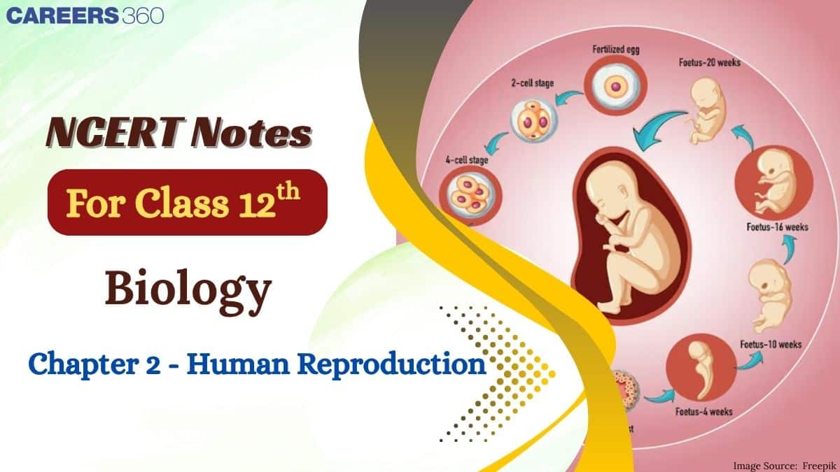
NCERT Class 12 Biology Chapter 3 Notes Human Reproduction
Have you ever wondered how a single cell transforms into a complete human being? The NCERT Class 12 Biology Chapter 2 Notes Human Reproduction make the complex processes of reproduction easier to understand, which allows students to revise faster. The Human Reproduction chapter covers topics like gametogenesis, fertilisation, and embryonic development. These NCERT Notes present all the important points clearly, helping students perform well in both board exams as well as in competitive exams like NEET.
This Story also Contains
- NCERT Class 12 Biology Chapter 2 Human Reproduction: Download PDF
- Class 12 Biology Chapter 2 Human Reproduction Notes
- Chapter 2 Human Reproduction: Previous Year Question and Answers
- How to Use NCERT Class 12 Biology Chapter 2 Notes Effectively?
- Advantages of Class 12 Biology Chapter 2 Human Reproduction Notes
- Chapter-Wise NCERT Class 12 Notes Biology
The NCERT Class 12 Biology Chapter 2 Notes also explain hormonal regulation, the menstrual cycle, and the process of parturition and lactation in a detailed manner. Diagrams and flowcharts are included to make complex processes easier to visualise and remember. Each topic is broken down into clear, short points for better understanding. These NCERT Notes for Class 12 highlight concepts in a structured way, making it easier to connect different processes, and act as a valuable resource for quick and effective revision.
Note: This chapter is renumbered as Chapter 2 in the NCERT Class 12 Biology textbook as per the latest syllabus.
NCERT Class 12 Biology Chapter 2 Human Reproduction: Download PDF
The Human Reproduction chapter covers the structure and function of male and female reproductive systems, gamete formation, and embryonic development. The NCERT Class 12 Biology Chapter 2 Notes present these topics in a simple and organised format for easy revision. Students can download the PDF and use it offline, making it convenient for anytime study. Regular study of the NCERT Notes for Class 12 Biology helps students strengthen their understanding and perform better in exams.
Also Read:
Class 12 Biology Chapter 2 Human Reproduction Notes
The Human Reproduction chapter is made easier to understand by breaking the complex process into simple steps. It covers topics like gametogenesis, fertilisation, implantation, and embryonic development, along with the hormonal regulation of the menstrual cycle. The chapter explains how these processes ensure the continuation of human life and the development of a healthy embryo. The notes present all important points of NCERT clearly, allowing students to revise the chapter quickly without missing any concepts.
Introduction to Reproduction
We know that humans are sexually reproducing and viviparous. The reproductive events in humans include the formation of gametes called gametogenesis, i.e., when the sperm in males transfer into the female genital tract, and fusion with the female gametes, which is called fertilisation, leads to the formation of a zygote. This is followed by the formation and development of the blastocyst and its attachment to the uterine wall, which is called implantation; the gestation period, in which embryonic development takes place; and finally, the delivery of the baby, which is parturition.
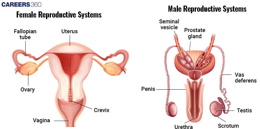
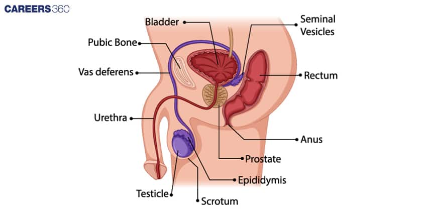
Male Reproductive System
The male reproductive system consists of the following organs:
1. | Primary sex organs | a pair of testes suspended in a scrotum |
2. | Secondary sex organs | The secondary sex organs include a system of ducts such as the rete testis, vasa efferentia, epididymis, vas deferens, and ejaculatory duct, along with accessory glands. |
3. | External genitalia | the penis, urethra, and scrotum |
Testes:
Situated outside the abdominal cavity in a pouch called the scrotum, which helps in maintaining the low temperature of the testes necessary for the process called spermatogenesis.
Each testis has around 250 testicular lobules, and each of them contains highly coiled seminiferous tubules in which sperm are produced. Each seminiferous tubule is lined by two types of cells, spermatogonia (male germ cells) and Sertoli cells.
Leydig cells:
It is also called the Interstitial cells. It is present around the seminiferous tubules that synthesize and secrete the hormone called androgen.
Ejaculatory duct:
Its work is to store and transport the sperm from the testes to the outside through the urethra. The urethra originates from the urinary bladder and extends through the penis to open at the tip.
Penis:
It is the male external genitalia. The enlarged end of the penis is called the glans penis, is covered by a loose fold of skin called the foreskin.
Male accessory glands
The male accessory ducts include:
paired seminal vesicles
prostate
paired bulbourethral glands
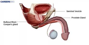
Secretion of these glands forms the seminal plasma, which contains fructose, calcium, and enzymes, and the secretion of bulbourethral glands also helps in the lubrication of the penis.
Female Reproductive System
Given below are the organs that together form the female reproductive system.
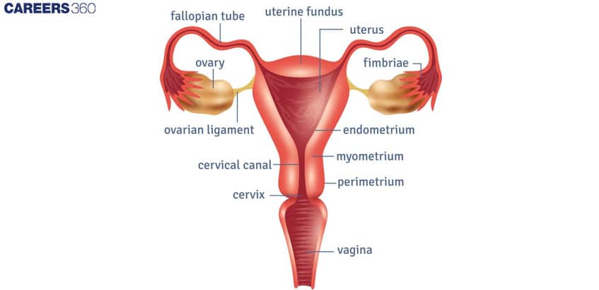
1. | Primary sex organ | a pair of ovaries |
2. | Secondary sex organ | duct system consisting of a pair of fallopian tubes, a uterus, cervix, and vagina |
3. | External genitalia | the opening of the birth canal, labia majora and labia minora, and the clitoris |
4. | Mammary glands | located in the breasts and responsible for lactation |
Ovaries:
They are the primary female sex organ that produces female gametes, eggs, and several other steroid hormones. Each ovary is covered by a thin epithelium that encloses the ovarian stroma, which is divided into two parts: a peripheral cortex and an inner medulla.
Fallopian tube:
The fallopian tube extends from the periphery of the ovary to the uterus. The part closer to the ovary is a funnel-shaped structure called the infundibulum, and it also has a finger-like projection called fimbriae.
The infundibulum leads to the ampulla and joins with the uterus through the isthmus. The uterus is pear-shaped in structure and is also called the womb.
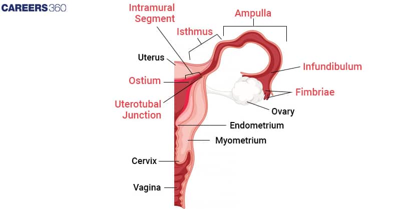
Uterus:
It opens the vagina through a narrow cervix. The cavity of the cervix (cervical canal), along with the vagina, forms the birth canal.
The wall of the uterus has three layers of tissue:
Perimetrium: The outermost thin layer that covers the external surface of the uterus.
Myometrium: The middle thick layer of smooth muscle that shows strong contractions during childbirth.
Endometrium: The innermost lining of the uterus that undergoes cyclic changes during the menstrual cycle to prepare for possible implantation.
Female external genitalia
Female external genitalia includes:
Mons pubis- It is a cushion of fatty tissue covered by skin and pubic hair.
Labia majora- It is a fleshy fold that surrounds the vaginal opening.
Labia minora- It is a paired fold of tissue under the labia majora.
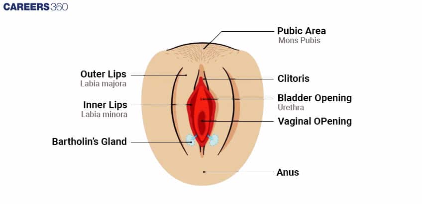
The opening of the vagina is often partially covered by a membrane called the hymen. The tiny finger-like projection present at the upper junction of the two labia minora above the urethral opening is called the clitoris.
Mammary glands
They are paired structures that contain glandular tissues and variable fats. Each glandular tissue contains 15-20 mammary lobes containing alveoli that secrete milk. Mammary ducts join to form the mammary ampulla.
Gametogenesis
It is the process of formation of male and female gametes in the testes and the ovary, respectively, is called gametogenesis.
Gametogenesis is of two types:
Spermatogenesis- In males
Oogenesis- In females
Spermatogenesis
It is the process through which sperm formation in males takes place. When testes are immature, male germ cells (spermatogonia) produce sperm that begin at the stage of puberty.
The spermatogonia present at the inner side of the seminiferous tubules multiply by mitotic division and increase in number. Each spermatogonium contains a total of 46 chromosomes, and then spermatogonia form a primary spermatocyte that undergoes meiosis to produce the secondary spermatocytes having 23 chromosomes.
The spermatids are transformed into spermatozoa by the process called spermiogenesis. The sperm heads remain embedded in Sertoli cells and are released from seminiferous tubules by the process of spermiation.
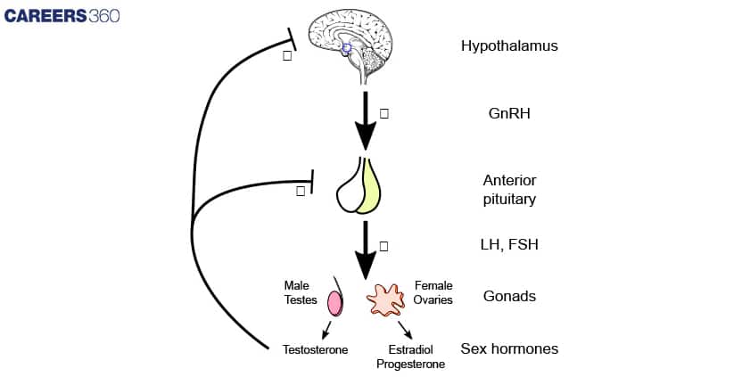
Hormonal control of spermatogenesis
Spermatogenesis is initiated due to an increase in the secretion of Gonadotropin-Releasing Hormone (GnRH) by the hypothalamus.
An increase in GnRH acts on the anterior pituitary and stimulates the secretion of two gonadotropins, LH and FSH.
LH acts on Leydig cells and stimulates them to secrete androgens.
FSH acts on Sertoli cells, stimulating the secretion of some factors that help in spermiogenesis.
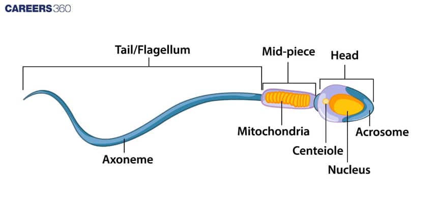
Structure of Sperm
Sperm is a microscopic structure composed of a head, neck, middle piece, and tail. The sperm head contains an elongated haploid nucleus, the anterior portion of which is covered by a cap-like structure acrosome.
A human male ejaculates about 200-300 million sperm during coitus. The seminal plasma, along with the sperm, constitutes the semen. The function of the male sex secondary ducts and glands is maintained by androgen hormones.
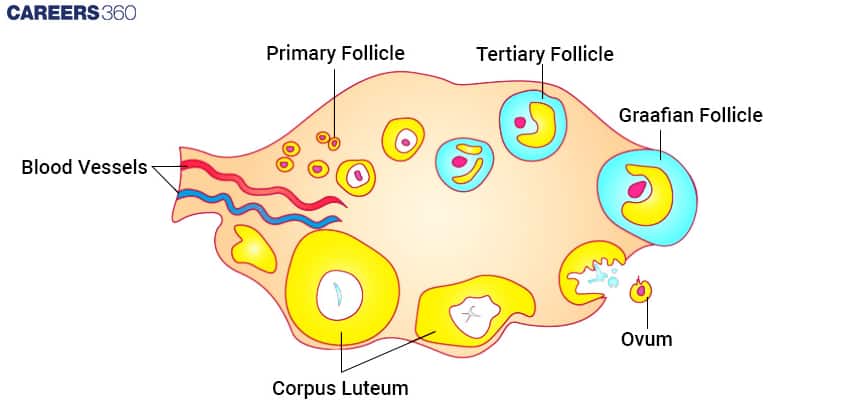
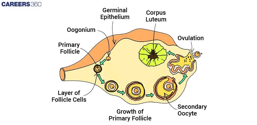
Oogenesis
It is the process of formation of mature female gametes is called oogenesis. It started during the embryonic development stage when millions of oogonia (gamete mother cells) are formed in each fetal ovary.
The gamete mother cells start division and enter prophase I of meiotic division, and are temporarily arrested at that stage, called primary oocytes.
Each primary oocyte is surrounded by granulosa cells, forming the primary follicle. At puberty, about 60,000- 80,000 primary follicles are left in each ovary.
The primary follicle gets surrounded by more layers of granulosa cells called secondary follicles that transform into tertiary follicles that contain fluid-filled cavities called the antrum.
The tertiary follicles further change into the mature follicle called the Graafian follicle, which ruptures to release secondary oocytes (ovum) from the ovary by the process of ovulation.
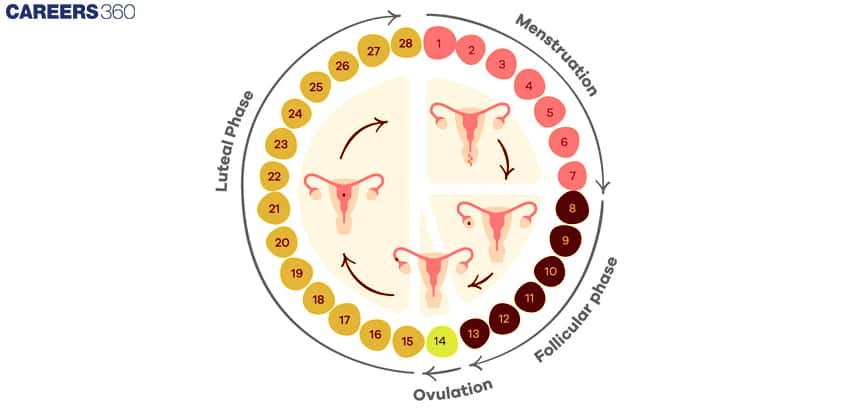
Menstrual cycle
The reproductive cycle in female primates is called the menstrual cycle. It starts at puberty and is called menarche.
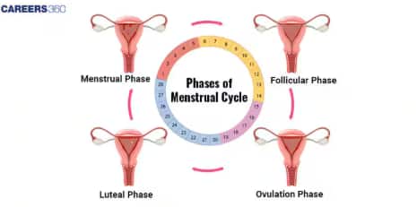
There are Four Phases of the Menstrual Cycle (MC):
Menstrual Phase:
In a 28-day menstrual cycle, the menses take place on a cycle of 3-5 days.
The production of LH from the anterior lobe of the pituitary gland is reduced.
The withdrawal of this hormone causes degeneration of the corpus luteum, and, therefore, progesterone production is reduced.
Production of oestrogen is also reduced in this phase.
The endometrium of the uterus breaks down & menstruation begins.
The cells of endometrium secretions, blood & unfertilised ovum constitute the menstrual flow.
Follicular Phase:
This phase usually includes cycle days 6-13 or 14 in a 28-day cycle.
The follicle-stimulating hormone (FSH) secreted by the anterior lobe of the pituitary gland stimulates the ovarian follicle to secrete oestrogens.
Oestrogen stimulates the proliferation of the endometrium of the uterine wall.
The endometrium becomes thicker by rapid cell multiplication, and this is accompanied by an increase in uterine glands & blood vessels.
Ovulatory Phase:
Both LH & FSH attain a peak level in the middle of the cycle (about the 14th day).
Oestrogen concentration in the blood increases.
Rapid secretion of LH induces the rupture of the Graafian follicle and thereby the release of the ovum.
In fact, LH causes ovulation.
Luteal Phase:
Includes cycle days 15 to 28.
Corpus luteum secretes progesterone.
Endometrium thickens.
Uterine glands become secretory.
Hormonal Control of Menstrual Cycle:
(i) FSH stimulates the ovarian follicles to produce oestrogens.
(ii) LH stimulates the corpus luteum to secrete progesterone.
(iii) The menstrual phase begins due to the fall in progesterone and estrogen levels, following degeneration of the corpus luteum.
(iv) LH causes ovulation
(v) The Proliferative phase is caused by the increased production of oestrogens.
(vi) The secretory phase is caused by increased production of progesterone.
Fertilisation and Implantation
The process of fertilisation and implantation is explained below:
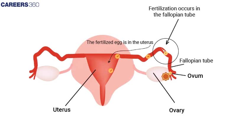
The process of fusion of sperm with an ovum is called fertilisation.
During coitus (copulation), semen is released into the vagina. The motile sperm swim rapidly to reach the junction of the isthmus and ampulla of the fallopian tube. The ovum also reaches there, and fusion of gametes takes place at the ampullary-isthmic junction.
The acrosome of sperm undergoes an acrosomal reaction and releases certain sperm lysins, which dissolve the egg envelopes locally and make a path for the penetration of sperm.
These sperm lysins contain a lysing enzyme. hyaluronidase, which dissolves the hyaluronic acid polymers in the intercellular spaces that hold the granulosa cells of corona radiata together; corona penetrating enzyme that dissolves the corona radiata, and acrosin, which dissolves the zona pellucida. Then it dissolves the zona pellucida.
Cortical reaction
(a) Immediately after the entry of sperm into the egg, the latter shows a cortical reaction to check the entry of more sperm.
(b) In this reaction, the cortical granules present beneath the egg’s plasma membrane release chemical substances between the ooplasm and the plasma membrane vitelline membrane.
(c) These substances raise the vitelline membrane above the egg surface. The elevated vitelline membrane is called the fertilization membrane.
(d) The increased space between the ooplasm and the fertilization membrane and the chemicals present in it effectively check the entry of other sperm.
(e) If polyspermy occurs, that is, more than one sperm enters the secondary oocyte, the resulting cell has too much genetic material to develop normally
The haploid gametes fuse together to form a diploid zygote. As the zygote moves towards the uterus, the mitotic division starts and forms cleavage to change into 2, 4,8,16 celled blastomeres.
The blastomeres with 8 to 16 cells are called the morula. Morula divides to change into blastocysts.
The blastomeres in the blastocyst are arranged into an outer layer called the trophoblast and an inner group of cells attached to the trophoblast called the inner cell mass.
The outer layer of the blastocyst is called the trophoblast, which attaches to the endometrium of the uterus, called implantation, which leads to pregnancy.
Pregnancy and embryonic development:
The finger-like projections on trophoblasts after implantation are called chorionic villi that, along with the uterine wall, form a functional unit between the developing embryo and maternal body called the placenta.
The placenta is attached to the fetus with an umbilical cord that transports food and oxygen to the embryo.
Hormones hCG (human chorionic gonadotropin)
hPL (human placental lactogen)
Relaxin
They are produced in women only during pregnancy by the placenta.
After implantation, the inner cell mass embryo differentiates into an outer layer called ectoderm and an inner layer called endoderm. A mesoderm soon appears between the ectoderm and the endoderm.
These three layers give rise to all tissues; organs in adults. It is important to note that the inner cell mass contains certain cells called stem cells, which have the potency to give rise to all the tissues and organs.
In humans, after one month of pregnancy, the embryo’s heart is formed. By the end of, 2nd month, limbs and digits are formed. By the end of the third month, major organs and external genital organs are well developed.
The first movement of the foetus is observed in 5 months. By the end of 24 weeks, the body is covered with fine hair, and eyelids and eyelashes are formed. At the end of 9 months fetus is fully developed.
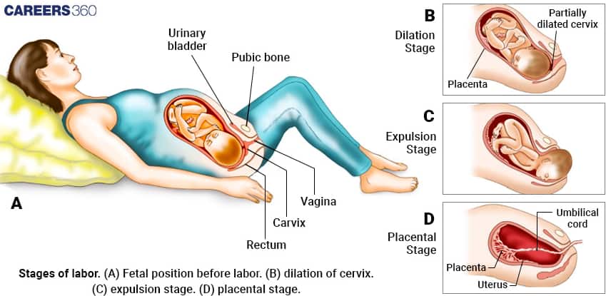
Parturition And Lactation
- Parturition is the process of delivery of a fully developed foetus is called parturition.
- Signals for parturition originate from the fully developed fetus and placenta, inducing mild uterine contractions called the foetal ejection reflex.
- It triggers the release of oxytocin from the maternal pituitary.
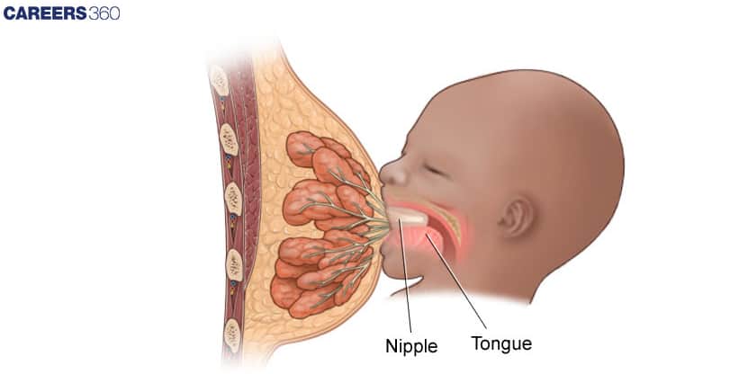
- The mammary glands of females start producing milk at the end of pregnancy through the process of lactation.
- The milk produced during the initial few days of lactation is called colostrum. Colostrum is rich in IgA antibodies that provide passive immunity to the newborn.
Also, Read
Confused between CGPA and Percentage?
Get your results instantly with our calculator!
Chapter 2 Human Reproduction: Previous Year Question and Answers
Some of the questions that have come in past years from the chapter are given below. Students will know how reproduction takes place in humans by referring to the Human Reproduction Class 12 Notes.
Question 1. Which of the following is not a component of the Fallopian tube?
Option 1. Uterine fundus
Option 2. Isthmus
Option 3. Infundibulum
Option 4. Ampulla
Answer :
The correct answer is option (1) as the uterine fundus is the upper, dome-shaped part of the uterus, above the opening of the fallopian tubes.
• Option (2) is incorrect as the isthmus is the last and narrow part of the oviduct that links to the uterus.
• Option (3) is incorrect as the infundibulum is the part of the oviduct that is closer to the ovary.
• Option (4) is incorrect as the ampulla is the wider part of the oviduct.
Hence, the correct answer is option (1). Uterine Fundus
Question 2. The Fallopian Tube provides an ideal environment for fertilization due to its:
Option 1. High acidity
Option 2. Thick mucus secretion
Option 3. Presence of spermicidal substances
Option 4. Optimal pH and supportive secretions
Answer :
The fallopian tube maintains a slightly alkaline pH, which helps sperm survive and move effectively. Its secretions contain nutrients and factors that support both sperm viability and egg fertilization. This favorable environment ensures successful fusion of gametes.
Hence, the correct answer is option (4), Optimal pH and supportive secretions
Question 3. In terms of their layered structures, which layers are shared between the allantois and the yolk sac?
Option 1. Outer Mesoderm and Inner Endoderm
Option 2. Outer Endoderm and Inner Mesoderm
Option 3. Outer Endoderm and Inner Ectoderm
Option 4. Outer Endoderm and Inner Endoderm
Answer :
Both the allantois and yolk sac are extraembryonic membranes that share a similar structure, consisting of an outer mesodermal layer and an inner endodermal lining. This arrangement helps in their role in nutrition, waste removal, and exchange during early embryonic development.
Hence, the correct answer is option (1), Outer Mesoderm and Inner Endoderm
Question 4. Which hormone is responsible for the release of the ovum from the ovary during the menstrual cycle?
Option 1. Estrogen
Option 2. Progesterone
Option 3. Luteinizing hormone (LH)
Option 4. Follicle-stimulating hormone (FSH)
Answer :
Ovulation is triggered by a sudden surge of luteinizing hormone (LH) from the anterior pituitary gland. This LH surge causes the mature Graafian follicle to rupture and release the ovum.
Hence, the correct answer is option (3), Luteinizing hormone (LH)
Question 5. The function of Sertoli cells in the seminiferous tubules is to
Option 1. Produce testosterone
Option 2. Nourish developing sperm
Option 3. Transport sperms
Option 4. Store sperms
Answer :
Sertoli cells provide nourishment and structural support to developing spermatogenic cells during spermatogenesis. They also help regulate the process of sperm formation.
Hence, the correct answer is option (2), nourish developing sperm
Also Read:
How to Use NCERT Class 12 Biology Chapter 2 Notes Effectively?
Understanding Human Reproduction is a bit difficult sometimes, but using notes in a smart and structured way makes the process much easier. A planned approach helps you grasp key concepts quickly and retain them for exams.
- Start with the male and female reproductive systems and their functions step by step from the Class 12 Biology Chapter 2 Human Reproduction Notes.
Revise the process of spermatogenesis and oogenesis using flowcharts, as they make the stages easy to understand.
Go through the hormonal control of reproduction, especially the menstrual cycle, with the help of diagrams showing FSH, LH, estrogen, and progesterone.
Highlight topics like fertilisation, pregnancy, and parturition, as questions are frequently asked about these concepts in the exam.
Use diagrams and flowcharts in the Class 12 Biology Chapter 2 Human Reproduction Notes for quick last-minute revision.
Advantages of Class 12 Biology Chapter 2 Human Reproduction Notes
Human Reproduction is an important chapter that explains how humans reproduce, grow, and develop. Studying this chapter through well-structured notes helps students understand complex processes easily.
- Class 12 Biology Chapter 1 Human Reproduction Notes PDF helps in understanding the structure and functions of male and female reproductive organs.
- The notes also explain gamete formation, fertilisation, and embryonic development in a detailed and clear manner.
- Studying through the notes makes it easier for students to answer diagram-based and application-based questions.
- Going through the notes regularly is useful for board exams as well as competitive exams like NEET.
- Class 12 Biology Chapter 1 Human Reproduction Notes PDF saves time during revision by explaining concepts in a simple and organised manner.
Chapter-Wise NCERT Class 12 Notes Biology
The following notes cover all chapters, including concepts, important definitions, and diagrams. They are useful for quick revision before exams and for strengthening understanding of the subject.
| NCERT Class 12 Biology Chapter 1 Sexual Reproduction in Flowering Plants Notes |
| NCERT Class 12 Biology Chapter 2 Human Reproduction notes |
| NCERT Class 12 Biology Chapter 3 Reproductive Health Notes |
| NCERT Class 12 Biology Chapter 4 Principles of Inheritance and Variation Notes |
| NCERT Class 12 Biology Chapter 5 Molecular Basis of Inheritance Notes |
| NCERT Class 12 Biology Chapter 6 Evolution Notes |
| NCERT Class 12 Biology Chapter 7 Human Health and Disease Notes |
| NCERT Class 12 Biology Chapter 8 Microbes in Human Welfare Notes |
| NCERT Class 12 Biology Chapter 9 Biotechnology: Principles and Processes Notes |
| NCERT Class 12 Biology Chapter 10 Biotechnology and its Applications Notes |
| NCERT Class 12 Biology Chapter 11 Organisms and Populations Notes |
| NCERT Class 12 Biology Chapter 12 Ecosystem Notes |
| NCERT Class 12 Biology Chapter 13 Biodiversity and Conservation Notes |
Frequently Asked Questions (FAQs)
Fertilisation in humans usually occurs in the ampullary–isthmic junction of the fallopian tube, where the sperm fuses with the ovum.
The placenta helps in the exchange of nutrients, oxygen, and wastes between the mother and the developing foetus and also secretes important hormones.
The Human Reproduction chapter is important because it covers fundamental concepts of reproduction, hormonal regulation, fertilisation, and embryonic development, which are frequently asked in board exams. These topics are also high-yield for competitive exams like NEET, making it essential to understand and revise them thoroughly.
In the NCERT Class 12 Biology Chapter 2 Notes Human Reproduction, you will study that the reproductive cycle in female primates is called the menstrual cycle and which starts at puberty and is called menarche, and occurs after every 28-30 days till the age of menopause.
Yes, oogenesis is the process of formation of mature female gametes is called oogenesis. The NCERT Class 12 Biology Chapter 2 Notes Human Reproduction say that it gets started during the embryonic development stage when millions of oogonia (gamete mother cells) are formed in each fetal ovary.
The main parts of the female reproductive system include the ovaries (produce eggs and hormones), fallopian tubes (transport eggs to the uterus), uterus (supports fetal development), cervix (which connects the uterus to the vagina), vagina (birth canal and menstrual passage), and external genitalia (vulva).
It is the process of formation of male and female gametes in testes and ovary respectively is called gametogenesis. Gametogenesis is of two types:
Spermatogenesis- Occurs in males
Oogenesis- Occurs in females

Popular Questions
A block of mass 0.50 kg is moving with a speed of 2.00 ms-1 on a smooth surface. It strikes another mass of 1.00 kg and then they move together as a single body. The energy loss during the collision is
| Option 1)
|
Option 2)
|
| Option 3)
|
Option 4)
|
An athlete in the olympic games covers a distance of 100 m in 10 s. His kinetic energy can be estimated to be in the range
| Option 1)
|
Option 2)
|
| Option 3)
|
Option 4)
|
A particle is projected at 600 to the horizontal with a kinetic energy . The kinetic energy at the highest point
| Option 1)
|
Option 2)
|
| Option 3)
|
Option 4)
|
In the reaction,
| Option 1)
|
Option 2)
|
| Option 3)
|
Option 4)
|
How many moles of magnesium phosphate, will contain 0.25 mole of oxygen atoms?
| Option 1)
0.02 |
Option 2)
3.125 × 10⁻² |
| Option 3)
1.25 × 10⁻² |
Option 4)
2.5 × 10⁻² |