

How Many Embryo Sacs are Present in an Ovule
Introduction
Each ovule contains a single embryo sac. The female gametophyte, commonly known as the embryo sac, is an oval structure present in the ovule of flowering plants. Each ovule contains one or more embryo sacs. Each ovary has a varied amount of ovules, which varies between plants. Maize, for example, can have a single seed or a vast number of them.
Ovule
The ovule is a component of seed plants' female reproductive system. It is the site of the production and maintenance of female reproductive components. Ovules are contained at the base of the carpel in ovaries, which has an aperture at the top, a stigma, and a style neck. Following fertilisation, these cells mature into a seed, which then ripens to become a fully formed adult plant. In flowering plants, the ovule is also known as the megasporangium.
Following fertilisation, the ovule develops and its wall begins to thicken in preparation for transformation into a seed. The ovary, on the other hand, begins to expand around it and develop into the fruit. Remember that certain plants, like the avocado, only have a single ovule in their ovary, whilst others, like the kiwifruit, have multiple ovules that eventually mature into multiple seeds in the fruit.
Confused between CGPA and Percentage?
Get your results instantly with our calculator!
Functions of Ovule
It is essential in the process of sexual reproduction.
Furthermore, it sends a pollen tube down through the style. The tube then penetrates the ovary and eventually reaches the plant's ovule.
The fertilisation process can then begin as the nucleus of the pollen grain is transported down the tube to merge with the nucleus of the embryo sac.
Notably, the male counterpart is usually pollen. It is home to the male gametophytes.
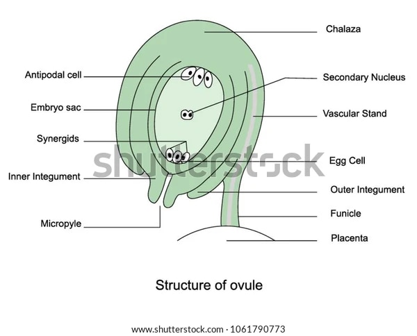
Structure of Ovule
The ovule is a megasporangium with an integument. It houses both the stalk and the body. A funicle is a name given to the stalk. The funicle has an attachment with the placenta on one end and an attachment with the body on the other.
However, the hilum is the place at which the funicle and the body connect. The funicle frequently fuses with the ovule's body on one side, forming a ridge known as a raphe.
The ovule's body contains two ends: the basal end and the upper end, also known as the micropylar end.
The primary body of the ovule is protected by one or two envelopes known as integuments. These leave a hole at the top of the ovule, which we call a micropyle. The integuments envelop a massive parenchymatous tissue known as the nucellus.
Parts and Develpoment of Ovule
Ovule orientation can be anatropous, as when the micropyle faces the placenta when inverted, campylotropous, amphitropous, or orthotropous. With the integuments that surround it, it seems to be a megasporangium.
The megaspores remain within them and divide through mitosis to produce the haploid female gametophyte or megagametophyte, which also remains within them.
Archegonia, which produce egg cells, are produced by gametophytes. It has a diploid zygote following the fertilisation process, and then cell division begins. Finally, the sporophyte embryo begins to develop. A second sperm nucleus unites with other nuclei in the megagametophyte to generate a polypoid endosperm tissue in flowering plants.
Types of Ovule
Ovules have been classified into six classes based on their morphologies:
Amphitropous: The ovule's body is so curled that both the ovule and the embryo sac resemble a horseshoe.
Orthotropous: The ovule body is straight here, allowing the micropyle, the chalaza, and the funicle that connects the ovule to the placenta, where the integuments and nucellus unite, to be aligned.
Anatropous: In this case, the ovules inverted during development, causing the micropyle to be situated near the hilum. A hilum scar marks the location of the funicle that attached the seed to the fruit wall.
Hemi-anatropous: Because their bodies develop at right angles to the funicle, these ovules appear to be lying on their side.
Circinotropous: The funicle in this case is unusually lengthy, making a nearly full circle around the ovule, whose micropyle eventually points upward.
Campylotropous: The micropyle and chalaza's synchronisation is broken, and the body is bent.
Female Gametophyte
The female gametophyte organ is commonly referred to as the Embryo sac. The female reproductive organ in gymnosperms is relatively big and multicellular, as the structure not only sustains the gametes but also aids in embryo development.
In angiosperms, on the other hand, the female gametophyte is a tiny, eight-nucleated structure that solely operates the gametes. Female gametophytes produce female gametes, which serve as the molecular foundation for fertilisation and aid in seed formation. Because this mechanism is supported by both gene and cellular processes, accessory cells can be activated genetically if gametes fail.
Development of Female Gametophyte
The development of female gametophytes occurs in two distinct stages. Megasporogenesis is the process by which tetrad haploid megaspores develop from a single diploid cell during meiosis. Only one megaspore survives and begins to develop inside an embryo sac; the other three die.
The operative haploid megaspore creates the female gametophyte-embryo sac during the second phase, megagametogenesis. The female gametophyte is composed of seven cells and eight nuclei. Only the polar nuclei of these eight nuclei shift and unite to form a diploid cell in the centre. This diploid cell merges with the sperm to form a triploid endosperm.
Embryo Sac
The female gametophyte, commonly known as the embryo sac, is an oval structure present in the ovule of flowering plants. When the haploid megaspore nucleus divides, an embryo sac is thought to form. It consists of two haploid nuclei and six haploid cells that lack cell walls. Sometimes the two haploid, polar nuclei merge to generate a single endosperm mother cell.
During fertilisation, one male nucleus unites with one egg nucleus to form a zygote, which grows into the embryo. When the 2nd male nucleus fuses with the primary endosperm nucleus, the endosperm nucleus is created. When this separates, the endosperm is created. In this article, we'll look at the construction and function of several structures created in the embryo sac.
Structure of Embryo Sac
The three cells at the micropylar end make form the egg apparatus, which consists of the egg and two synergids. Three antipodal cells can be found at the opposite end of the embryo sac. The massive central cell, which sits between the two groups of cells, contains two polar nuclei, one from each of the two groups of four nuclei.
When the two polar nuclei eventually combine and fuse, a diploid secondary nucleus is formed. As a result, the mature embryo sac has seven cells. All of the cells in the embryo sac are haploid except for the centre cell, which is diploid (due to the fusion of two polar nuclei).
Classification of Embryo Sac
Embryo sacs are classified into three categories of megaspores:
Monosporic: The nucellus meiosis of the diploid megaspore mother cell produces a Polygonum-type embryo sac or four haploid megaspores in the monosporic. Three megaspores, usually those near the nucellus' micropylar end, die as a result of programmed cell death, leaving only one working megaspore.
Diasporic: Meiosis produces just two megaspores, each with two haploid nuclei because there is no cytokinesis or cell plate growth after the second meiotic division in diasporic embryo sacs. The megaspore nearest to the micropyle then dies, leaving only one working megaspore with two haploid nuclei.
Tetrasporic: Because cell plates fail to form during both meiotic divisions, tetrasporic embryo sacs have a single four-nucleate megaspore.
Embryo Sac Formation
The process through which the female gametophyte matures in plants, during which the megaspore arises from megasporogenesis and grows into the embryo sac, which habitats the female gamete, is known as mega gametogenesis.
The growing embryo goes through meiosis, which has multiple stages, during the process of megasporogenesis. The surviving megaspore undergoes mitosis where each nucleus migrates to opposing ends of the embryo sac while producing a structure with a binucleate embryo sac.
The haploid nucleus undergoes two rounds of mitosis, resulting in four haploid nuclei on either end of the embryo sac. The embryo sac's nuclei migrate to the centre to form the binucleate endosperm mother cell, leaving three nuclei on the micropylar end.
Conclusion
The ovary is the bigger basal component of the pistil, a flower's female organ, in botany. Ovules are found in the ovary and grow into seeds after fertilisation. The ovary will develop into a fruit, either dry or fleshy, containing the seeds.
A mature ovule is made up of a food tissue surrounded by one or two future seed coats called integuments. A small aperture in the integuments (the micropyle) allows the pollen tube to enter and discharge its sperm nuclei into the embryo sac, a big oval cell where fertilisation and development take place. Each ovule is linked to the stalk (funiculus) by its base.
The embryo sac develops normally from the bottom of a row of four axial megaspores. The synergids are very lengthy, and the egg is sandwiched between them. The pollen tube connects the synergids, and the union of the male and egg nuclei was detected.
